Review sheet 30 anatomy of the heart – As Review Sheet 30: Anatomy of the Heart takes center stage, this opening passage beckons readers into a world crafted with precision and clarity, ensuring a reading experience that is both absorbing and distinctly original. Prepare to embark on a journey through the heart’s intricate structure, where each beat echoes the symphony of life.
This comprehensive guide unveils the heart’s external and internal anatomy, delving into its chambers, valves, and the intricate network of blood vessels that sustain our very existence. Histological insights illuminate the cellular building blocks of the heart, while embryological perspectives trace its remarkable developmental odyssey.
Overview of the Heart’s Anatomy

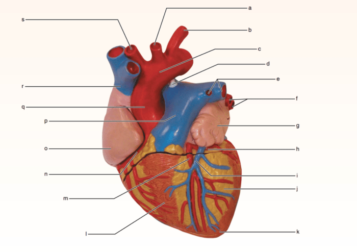
The heart is a muscular organ located in the thoracic cavity, medially between the lungs, and slightly shifted towards the left. It is enclosed within the pericardial sac, which provides protection and lubrication.
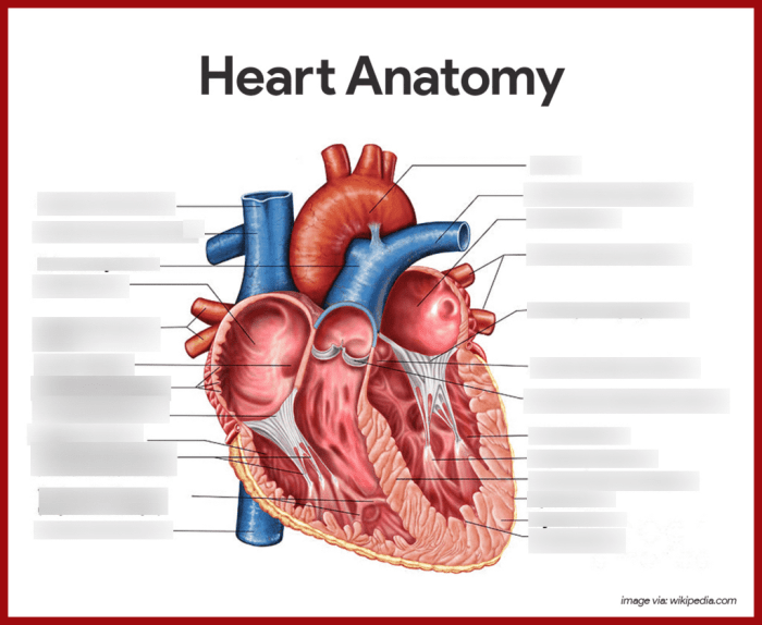
The heart has four chambers: two atria (right and left) and two ventricles (right and left). The atria receive blood from the body and lungs, while the ventricles pump blood out to the body and lungs.
The heart has four valves: the tricuspid valve, pulmonary valve, mitral valve, and aortic valve. These valves prevent backflow of blood within the heart.
The heart is supplied with blood by the coronary arteries and drained by the coronary veins.
Pericardial Sac
The pericardial sac is a double-walled sac that surrounds the heart. The outer layer is fibrous and provides structural support, while the inner layer is serous and produces pericardial fluid. The pericardial fluid reduces friction between the heart and the surrounding structures.
External Anatomy of the Heart: Review Sheet 30 Anatomy Of The Heart

The heart is a muscular organ located in the mediastinum, the middle compartment of the chest cavity. It is approximately the size of a clenched fist and is oriented with the apex (tip) pointing down and to the left, while the base (wider part) is directed upward and to the right.
Chambers of the Heart
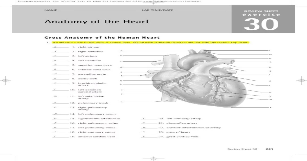
The heart consists of four chambers: two atria (singular: atrium) and two ventricles (singular: ventricle). The right atrium and right ventricle form the right side of the heart, while the left atrium and left ventricle form the left side.
- Right Atrium:Receives deoxygenated blood from the body via the superior and inferior vena cavae.
- Right Ventricle:Pumps deoxygenated blood to the lungs via the pulmonary artery.
- Left Atrium:Receives oxygenated blood from the lungs via the pulmonary veins.
- Left Ventricle:Pumps oxygenated blood to the body via the aorta.
Coronary Sulcus and Vessels
The coronary sulcus is a groove that encircles the heart, separating the atria from the ventricles. It contains the coronary vessels, which supply the heart muscle with oxygenated blood.
- Coronary Arteries:Branch off from the aorta and supply oxygenated blood to the heart muscle.
- Coronary Veins:Drain deoxygenated blood from the heart muscle and return it to the right atrium.
3. Internal Anatomy of the Heart
The internal anatomy of the heart consists of several structures, including the endocardium, myocardium, epicardium, heart valves, and chambers. These components work together to facilitate the efficient pumping of blood throughout the body.
Structure and Function of the Heart’s Layers, Review sheet 30 anatomy of the heart
The heart is composed of three distinct layers:
- Endocardium:The innermost layer, which lines the heart’s chambers and valves. It is composed of endothelial cells and provides a smooth surface for blood flow.
- Myocardium:The middle layer, which forms the muscular walls of the heart. It is responsible for the heart’s contractions and pumping action.
- Epicardium:The outermost layer, which covers the heart and is continuous with the parietal pericardium, a membrane that surrounds the heart.
Heart Valves
The heart valves are essential for maintaining the proper flow of blood through the heart. There are four main heart valves:
- Tricuspid valve:Located between the right atrium and right ventricle, it prevents blood from flowing back into the atrium during ventricular contraction.
- Pulmonary valve:Situated at the exit of the right ventricle, it prevents blood from flowing back into the ventricle during ventricular relaxation.
- Mitral valve (bicuspid valve):Located between the left atrium and left ventricle, it prevents blood from flowing back into the atrium during ventricular contraction.
- Aortic valve:Situated at the exit of the left ventricle, it prevents blood from flowing back into the ventricle during ventricular relaxation.
Flow of Blood through the Heart
The flow of blood through the heart is a continuous process that involves the coordinated action of the heart’s chambers and valves:
- Deoxygenated bloodfrom the body enters the right atrium through the superior and inferior vena cavae.
- The right atrium contracts, pushing blood through the tricuspid valve into the right ventricle.
- The right ventricle contracts, propelling blood through the pulmonary valve into the pulmonary artery, which carries it to the lungs for oxygenation.
- Oxygenated bloodfrom the lungs returns to the heart via the pulmonary veins and enters the left atrium.
- The left atrium contracts, forcing blood through the mitral valve into the left ventricle.
- The left ventricle contracts, pumping blood through the aortic valve into the aorta, the main artery that distributes blood to the body.
4. Histology of the Heart

The heart is a muscular organ responsible for pumping blood throughout the body. Its unique histological structure enables it to perform this vital function effectively.
Cardiac Muscle Cells
Cardiac muscle cells, also known as cardiomyocytes, are specialized cells that make up the bulk of the heart’s tissue. These cells are branched and interconnected, forming a syncytium that allows for coordinated contractions.
Each cardiomyocyte has a central nucleus and numerous mitochondria, reflecting its high energy demands. The cytoplasm contains myofibrils, composed of thick and thin filaments that slide past each other during contraction.
Intercalated Discs
Intercalated discs are specialized junctions between adjacent cardiomyocytes. They play a crucial role in coordinating the electrical and mechanical activity of the heart.
Intercalated discs consist of three main components: desmosomes, gap junctions, and fascia adherens. Desmosomes provide mechanical stability, while gap junctions allow for the rapid spread of electrical impulses, ensuring synchronized contractions.
Blood Vessels of the Heart
The heart has a rich network of blood vessels that supply it with oxygen and nutrients and remove waste products.
- Coronary arteries:These arteries branch off the aorta and supply oxygenated blood to the heart muscle.
- Coronary veins:These veins collect deoxygenated blood from the heart muscle and return it to the right atrium.
- Thebesian veins:These small veins drain blood directly from the heart muscle into the heart chambers.
5. Embryology of the Heart
The heart is one of the first organs to form during embryogenesis, beginning as a simple tube that eventually develops into the complex four-chambered structure.The development of the heart can be divided into several stages:
-
-*Cardiac crescent formation
The heart begins as a crescent-shaped structure on the ventral side of the embryo.
-*Linear heart tube formation
The cardiac crescent folds and fuses to form a linear heart tube.
-*Looping of the heart tube
The heart tube bends and loops to the right, forming the primitive atrium and ventricle.
-*Chamber formation
The atrium and ventricle are divided into two chambers each, forming the four-chambered heart.
-*Valve formation
The valves of the heart develop from swellings in the endocardial cushions, which are located at the junctions of the chambers.
Congenital heart defects are structural abnormalities of the heart that are present at birth. These defects can range from mild to severe and can affect any part of the heart. Some common congenital heart defects include:
-
-*Atrial septal defect (ASD)
A hole in the wall between the atria.
-*Ventricular septal defect (VSD)
A hole in the wall between the ventricles.
-*Tetralogy of Fallot
A combination of four heart defects, including a VSD, pulmonary stenosis, an overriding aorta, and right ventricular hypertrophy.
-*Coarctation of the aorta
A narrowing of the aorta, the main artery that carries blood away from the heart.
The embryological origins of congenital heart defects are not fully understood, but it is thought that they are caused by a combination of genetic and environmental factors.
6. Clinical Significance of Heart Anatomy
Understanding the intricate anatomy of the heart is paramount for healthcare professionals, as it provides the foundation for accurate diagnosis and effective treatment of cardiovascular diseases.Imaging techniques, such as echocardiography and cardiac MRI, play a crucial role in visualizing the heart’s anatomy non-invasively.
These techniques allow clinicians to assess cardiac structure, function, and blood flow patterns, aiding in the detection and characterization of various heart conditions.Moreover, a thorough understanding of heart anatomy is essential for surgical procedures. Cardiac transplantation, for instance, requires precise knowledge of the heart’s anatomical landmarks and vascular connections to ensure successful graft placement and minimize complications.
Similarly, valve replacement surgeries necessitate familiarity with the specific anatomy of the affected valve and its surrounding structures.
User Queries
What is the function of the pericardial sac?
The pericardial sac protects the heart and provides a lubricating fluid to reduce friction during contractions.
Which valve separates the right atrium from the right ventricle?
Tricuspid valve
What is the histological structure of cardiac muscle cells?
Cardiac muscle cells are branched, striated cells connected by intercalated discs, allowing for coordinated contractions.